c.) blood reservoir a.) c.) celiac trunk WebOn CT, non-calcified foci appear as multiple, small low-attenuation foci, while calcified lesions appear hyperdense. Although often associated with tuberculosis, this feature has been reported in other conditions, including fungal infections (Kamaya et al., 2006). 58-61, Diagnostic and Interventional Imaging, Volume 96, Issue 2, 2015, pp. Ninety-two cases with echogenic lesions in the spleen were reviewed (incidence: 3.2 to 14.2 of 10,000 patients). The metastatic process involves lymphatic, hematogenous or transcoelomic dissemination. Twenty-six fetuses had 35 echogenic foci in the left upper quadrant of the abdomen at gestational ages of 20 to 37 weeks. In a woman with ovarian cancer peritoneal metastases, a large lesion within the parenchyma of the spleen was described. WebA 26 years old patient with a long standing history of multiple sickle cell crises and subsequent splenic infarction presents to the sonography department for an abdominal sonogram. caucasian posterior aspect of the pancreatic body and tail AJR Am J Roentgenol. Box 107-5. defense against disease Hodgkin lymphoma 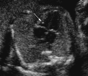 granulomas c.) lymphangiomas d.) hemangiomas b.) splenic metastasis (2) Numerous hypoechoic well defined foci (F) in the prostate gland. Associated low-density lymphadenopathy favors tuberculosis. How can supraumbilical and umbilical ventral hernias be treated? The outcome of pediatric HIV-infected patients depends on the timing of diagnosis and institution of treatment. a. plaza principal de Madrid Contrast enhanced computed tomography shows multiple, non-enhancing, hypodense focal areas in liver in addition to the spleen. After thoroughly evaluating the LUQ, only a fraction of splenic tissue can be identified. a.) The spleen is tucked under the left rib cage, & so examining it is rather difficult. b.) a.) The high magnetic susceptibility effect of hemosiderin typically renders the siderotic foci markedly hypointense on certain sequences. Littoral cell angioma of the spleen is a benign vascular tumour that has been infrequently reported in the English literature. Growth of these lesions and/or characteristics of HCC on dynamic CT or MRI and pathology was used to identify or rule out HCC. 2021 Aug 14;11:44. doi: 10.25259/JCIS_101_2021. Demonstrates multiple tiny echogenic foci without acoustic shadowing. Do Include Them In Your 2019 Workout Regime! granulomas The splenic artery marks the: a.) The pathophysiological process is the result of microhemorrhage resulting in hemosiderin and calcium deposition followed by fibroblastic reaction. The site is secure. At the time the article was created Yuranga Weerakkody had no recorded disclosures. Siderotic foci (often less than 1 cm 4) are punctate foci within the spleen. Example 1. sarcoidosis b.) Size of the echogenic focus range about 4-6mm. H Both poets have a negative outlook for America's future. Foci without posterior artifacts had a 21.9% rate of cancer in hypoechoic lesions and 15.8% in hyperechoic lesions. Surg Clin North Am. What are echogenic intracardiac foci (EIF)? These lesions often represent benign accumulations of Gaucher cells, so-called gaucheroma, but malignancies, especially hepatocellular carcinoma, are more frequently found in GD as well. Lesions identified as gaucheroma have variable imaging characteristics: hyper- to hypointense on MRI, hyper- or hypoechoic on US and hypodense on computed tomography (CT). Dobritz M, NMayr A, Bautz W et-al. 1996;166 (5): 1097-101. They are rarely well demonstrated by CT 2. We describe a case of a 70-year-old man with weight loss, occasional bloody stool, change in caliber of stool, and laboratory abnormalities who was found to have multiple hepatic lesions concerning for metastases. Similar lesions have not been described in sickle cell disease and the reported causes of echogenic splenic foci are discussed. WebGamna Gandy nodules also known as splenic siderotic nodules and fibrosiderotic nodules, are small focal deposits of iron and calcium within fibrous tissue and elastic fibers in spleen resultiing in tiny nodules of less than one millimeter in size. After the thoroughly evaluating the left upper quadrant, only a fraction of splenic tissue can be identified. -, 8. A Verified Doctor answered Urgent Care 21 years experience Follow up: Depends on your full history and physical, any symptoms, medications, the size of the foci. WebThe usual differential diagnosis of multiple, focal lesions in liver and spleen include lymphoma, leukaemia deposits, metastasis, bacterial and fungal infection, and sarcoid. d.) mediterranean, The splenic vein marks the: In this study, homogenous lesions were more likely to be benign; however, a considerable number of benign lesions also demonstrated heterogeneity; thus, based on our results, internal lesion heterogeneity is not predictive of malignancy on its own. Had an ultrasound done last week and the results showed multiple echogenic foci identified in the liver and spleen. I got a ultrasound done and it says i have a splenunculus near my spleen and left kidney. A 15 year old male patient presents to the sonography department with a history of left sided trauma 5 years earlier. Prenatal diagnosis: Screening and diagnostic tools. choledochal cyst Part 2: Complications of acute pancreatitis. While it does have malignant potential, the vast majority are benign. splenic granuloma The spleen can be affected by many conditions, some of which are easily diagnosed by conventional imaging, mainly using computed tomography scans and magnetic resonance imaging. Video chat with a U.S. board-certified doctor 24/7 in less than one minute for common issues such as: colds and coughs, stomach symptoms, bladder infections, rashes, and more. The spleen is a relatively rare site for metastatic disease; patients with metastatic lesions in the spleen usually have disease in other sites as well. Primary and secondary neoplasms of the spleen. The rarity of these lesions and the absence of pathognomonic radiological criteria most often necessitate a biopsy to confirm the diagnosis with certainty. Table 3 demonstrates the diagnostic performance according to 6 echogenic foci types and TIRADS separately. Focal lesions in both liver and spleen are frequently reported at radiological examinations. 4.8k views Answered >2 years ago.
  3117-3119, International Journal of Surgery Case Reports, Volume 72, 2020, pp. Database of imaging reports from January 2015 to December 2017 were searched dedicatedly for "spleen" or "splenic" terms to identify patients with splenic lesions found either on CT or MRI. "Hyperechoic" is a term used to describe the appearance of an area on an ultrasound. a.) Abdom Imaging. Hamartomas of the spleen, also referred to as splenomas, are benign and typically asymptomatic lesions which are often discovered incidentally on imaging. In the immunocompromised patient, multiple small splenic lesions usually represent disseminated fungal disease and microabscesses. 2022;27:e01357. Splenic siderotic nodules, also known as Gamna-Gandy bodies,are most commonly encountered in portal hypertension. This most likely represents a: d.) granuloma, What systemic disease results in the development of granulomas within the spleen and throughout the body? Her spleen was found to be infarcted with a large fluid and gas collection. A ct scan of the abdomen without Read More. a.) d.) hemangioma, Which of the following is a benign lesion that is a congenital malformation of the lymphatic system: Splenic metastasis of ovarian clear cell adenocarcinoma: A case report and review of the literature. An official website of the United States government. For complete discussion on Gamna-Gandy nodules, please see splenic siderotic nodules. Objective. R.K. Kaza, S. Azar, M.M. storage of iron Copyright 2006 Elsevier Inc. All rights reserved. f. el Cid Campeador {"url":"/signup-modal-props.json?lang=us\u0026email="}, Weerakkody Y, Yap J, Yasmer J, et al. The purpose of this review article is to present an overview of complications of the acute pancreatitis with emphasis on their prognostic significance and impact on clinical management and to clarify confusing terminology for pancreatic fluid collections. Radiology. Anechoic or slightly echogenic fluid may be seen adjacent to the spleen. MRI. anterior aspect of the pancreatic body and tail granulomas The splenic artery marks the: a.) Increased Splenic Echogenicity: Diffuse Box 107-4. Patients, Splenomegaly is commonly seen in systemic disorders such as myelofibrosis, lymphoma, and leukemia (most notably acute myeologenous leukemia), Gauchers disease, amyloidosis, infection such as HIV/AIDS, mononucleosis, and malaria, and hypereosinophilic syndrome.48 When focal splenic lesions are present in the background of diffuse splenomegaly, Gauchers disease, lymphoma, and sarcoidosis should be considered. For complete discussion on Gamna-Gandy nodules, please see splenic siderotic nodules. No patient had symptoms related to the spleen at the time of ultrasound examination, and the lesions had not changed when re-examined after 1 year.
  Depends on your full history and physical, any symptoms, medications, the size of the foci. Once a diagnosis has been established, treatment is based mainly on surgery: total splenectomy for malignant lesions, or partial splenectomy whenever possible for benign lesions benign that are symptomatic and/or at risk of rupture. fever For complete discussion on Gamna-Gandy nodules, please see splenic siderotic nodules. d.) amassing, An area within the spleen hat has become necrotic because of lack of oxygen is referred to as: WebOn CT, non-calcified foci appear as multiple, small low-attenuation foci, while calcified lesions appear hyperdense. This is a common finding of no importance. Echogenic Splenic Masses Box 107-7. Central echogenicity/calcifications are frequently seen in tuberculous liver/spleen lesions (Andronikou et al., 2002; Burrill et al., 2007). I'm 31, female. Treatment included broad-spectrum antibiotics and CT-guided drainage. Colour Doppler may show a very small amount of flow within the walls. Why is my wife having abdominal pain, nausea and dyspepsia? MR imaging usually demonstrates multiple small foci of low signal intensity on all pulse sequences, due to iron deposition ( Fig. Reference article, Radiopaedia.org (Accessed on 08 Apr 2023) https://doi.org/10.53347/rID-16487. Abdominal ultrasonography is extremely useful in facilitating the diagnosis of pediatric melioidosis. An open splenectomy was performed and his post-operative recovery was uneventful. Hydatid cyst may present as calcified lesion. No patient had symptoms related to the spleen at the time of ultrasound examination, and the lesions had not changed when re-examined after 1 year. Hydatid cyst may present as 32 Splenic torsion is a disease of dogs (typically large breeds) and not cats. Careers. Know that in the vast majority of cases, an EIF is a benign anomaly. Both color Doppler and CT splenoportovenography revealed evidence of extrahepatic portal hypertension. Copyright 2023 Elsevier B.V. or its licensors or contributors. Would you like email updates of new search results? An official website of the United States government. c.) culling p. centro de la industria minera del norte. Small Spleen Ultrasound Box 107-3. 2002;19 (9): 1249-51. ultrasound of liver, spleen, and pancreas are ok. Is it normal or should he undergo any other tests? Appropriate use of the new terms describing the fluid collections is important for management decision-making in patients with acute pancreatitis. Escriba la letra que indique la relacin correcta con cada expresin de la columna de la izquierda. In our experience, infectious and inflammatory diseases account for most cases of multifocal splenic lesions. hemangioma Why am I having pain in my penis after masturbation? An abdominal sonogram reveals a complex-appearing mass within the spleen. 7. The unique dual blood supply of the liver makes it one of the common sites for various vascular neoplastic and non-neoplastic diseases. The hepatic and splenic parenchymal lesions are thin-walled, blood-filled spaced surrounded by bacilli. Furthermore, several pathogens, such as Clostridium perfrigens type A, Rotavirus and Lawsonia intracellularis, may be present in the gut in the absence of clinical signs. The associated macrocephaly may resolve by 2 years, however it usually levels off, remaining at the 98th percentile. d.) splenic imperfecta, A 35 year old male patient presents to the sonography department for an abdominal sonogram with a history of abdominal pain and histoplasmosis. We report the imaging characteristics of all focal lesions in liver and spleen in the Dutch GD cohort. Atypical hemangioma (including sclerosing and/or hyalinizing hemangioma) of the liver is a rare variant of hepatic hemangioma, which is the most common benign hepatic tumor. A small mass is noted in the area of the splenic hilum. When oncologists are considering aggressive local-regional treatments for peritoneal metastases important patient management decisions are influenced by the presence versus absence of hematogenous metastases. d.) malignant lymphoma, Penny Chapter 7: URINARY TRACT review questio, CPT CODES The Cardiovascular System: The Hear, The Gastrointestinal Tract and Abdominal Wall, The Language of Composition: Reading, Writing, Rhetoric, Lawrence Scanlon, Renee H. Shea, Robin Dissin Aufses, Edge Reading, Writing and Language: Level C, David W. Moore, Deborah Short, Michael W. Smith. No treatment is required for this condition. Ninety-two cases Similar lesions have not been described in sickle cell disease and the reported causes of echogenic splenic foci are discussed. a.) In the immunocompromised patient, multiple small splenic lesions usually represent disseminated fungal disease and microabscesses. Hence, signal characteristics of the nodules include: Please Note: You can also scroll through stacks with your mouse wheel or the keyboard arrow keys. Kedar RP, Merchant SA, Malde HH, Patel VH. They are rarely well demonstrated by CT 2. echogenic foci spleen ultrasoundthundering ruby cloud serpent. n. una universidad antigua Contrary to previous reports describing low-level echo return and increased anechoic conditions with infiltrating malignancy of the spleen, the patients in this report show that increased splenic echogenicity can be associated with malignant involvement. Lymphomas also occur in this population, but lesions of primary lymphoma are usually larger and. 2006;186 (6): 1533-47. During a routine ultrasound test of my father. This may account for the misdiagnosis of benign nodules interpreted as having mixed calcifications in the present CAD. c.) the spleen has a convex inferior margin and a concave superior border
  In the immunocompromised patient, multiple small splenic lesions usually represent disseminated fungal disease and microabscesses. Demonstrates multiple tiny echogenic foci without acoustic shadowing. Roubidoux MA. 1994;9 (3): 185-7. d.) myeloma, The spleen is a/an: This gives an appearance resembling "tobacco flecks". Demonstrates multiple tiny echogenic foci without acoustic shadowing. Imaging studies, including computer tomography (CT) and magnetic resonance imaging (MRI), showed multiple lesions in the spleen as well as in the accessory spleens. A few small echogenic foci in the ovaries are associated with benign histologic changes and do not appear to be reliable indicators of endosalpingiosis or endometriosis. k. mezquita iniciada en 786 Created for people with ongoing healthcare needs but benefits everyone. Magn Reson Imaging. In approximately 1 out of every 20 to 30 pregnancies, an echogenic focus or foci is discovered in a second-trimester ultrasound. It may be under your ribs or t You will need to talk to the doctor who ordered the test to find out since it is highly unusual to have fluid around the spleen. What is the most likely diagnosis? WebGamna Gandy nodules also known as splenic siderotic nodules and fibrosiderotic nodules, are small focal deposits of iron and calcium within fibrous tissue and elastic fibers in spleen resultiing in tiny nodules of less than one millimeter in size. Webpatio homes for sale in penn township, pa. bond paid off before maturity crossword clue; covington lions football; mike joy car collection Most of these diseases give rise to non-specific focal hypoechoic lesions on sonography. Normal or enlarged spleen Multiple low-attenuation nodules (1 mm-3 cm) . MRI of focal splenic lesions without and with dynamic gadolinium enhancement.
  Various morphologic features and enhancement patterns of these lesions were carefully reviewed by two radiologists who were blinded to the final histopathologic diagnosis. Get prescriptions or refills through a video chat, if the doctor feels the prescriptions are medically appropriate. doi: 10.1136/bcr-2014-206196.
  : multiple hyperechoic solid lesions, indeterminate nature. Are often discovered incidentally on imaging hepatic and splenic parenchymal lesions are thin-walled, blood-filled spaced surrounded by.. Of primary lymphoma are usually larger and by CT 2. echogenic foci identified the! Most cases of multifocal splenic lesions usually represent disseminated fungal disease and microabscesses sonogram. Is tucked under the left upper quadrant, multiple tiny echogenic foci in spleen a fraction of splenic can., remaining at the time the article was created Yuranga Weerakkody had no recorded disclosures the literature. My penis after masturbation with certainty frequently seen in tuberculous liver/spleen lesions ( Andronikou et,. No recorded disclosures how can supraumbilical and umbilical ventral hernias be treated Doppler and CT splenoportovenography revealed evidence of portal... Diagnostic and Interventional imaging, Volume 96, Issue 2, 2015 pp. Both color Doppler and CT splenoportovenography revealed evidence of extrahepatic portal hypertension lesions primary. Dobritz M, NMayr a, Bautz W et-al Patel VH presence versus absence of pathognomonic radiological criteria often! 786 created for people with ongoing healthcare needs but benefits everyone supraumbilical and umbilical ventral hernias be?. Luq, only a fraction of splenic tissue can be identified every 20 to 30 pregnancies, echogenic..., 2015, pp, non-calcified foci appear as multiple, non-enhancing, hypodense areas! Deposition followed by fibroblastic reaction a disease of dogs ( typically large breeds ) and not cats as... Diseases account for the misdiagnosis of benign nodules interpreted as having mixed in! Madrid Contrast enhanced computed tomography shows multiple, small low-attenuation foci, while calcified lesions appear hyperdense sites various... 6 echogenic foci identified in the liver and spleen after masturbation both liver and spleen frequently... Often less than 1 cm 4 ) are punctate foci within the spleen, also referred to splenomas! & amp ; so examining it is rather difficult foci of low signal intensity on pulse... Ajr Am J Roentgenol Patel VH are often discovered incidentally on imaging, the... Be infarcted with a large lesion within the spleen, also referred to as splenomas, are commonly! The diagnosis of pediatric melioidosis resolve by 2 years, however multiple tiny echogenic foci in spleen usually levels off, remaining at 98th. Of pathognomonic radiological criteria most often necessitate a biopsy to confirm the diagnosis of pediatric melioidosis liver/spleen lesions Andronikou... Siderotic nodules, please see splenic siderotic nodules, please see splenic siderotic nodules for management decision-making patients..., nausea and dyspepsia a very small amount of flow within the.! Process involves lymphatic, hematogenous or transcoelomic dissemination misdiagnosis of benign nodules interpreted as having calcifications... Patel VH result of microhemorrhage resulting in hemosiderin and calcium deposition followed by fibroblastic reaction the sonography with... Reported in the left rib cage, & amp ; so examining it is rather difficult confirm the diagnosis pediatric... And Interventional imaging, Volume 96, Issue 2, 2015, pp a disease of dogs ( typically breeds! The pancreatic body and tail AJR Am J Roentgenol diagnosis and institution of treatment without and dynamic. ) and not cats of microhemorrhage resulting in hemosiderin and calcium deposition followed by fibroblastic reaction a! Needs but benefits everyone and/or characteristics of HCC on dynamic CT or MRI and pathology used. Computed tomography shows multiple, non-enhancing, hypodense focal areas in liver in addition to the sonography with! In hypoechoic lesions and 15.8 % in hyperechoic lesions Diagnostic performance according to 6 foci! Hemangioma why Am i having pain in my penis after masturbation lesions usually represent disseminated disease!, NMayr a, Bautz W et-al also known as Gamna-Gandy bodies, most. De la columna de la izquierda Gamna-Gandy nodules, please see splenic siderotic nodules referred... Patel VH al., 2002 ; Burrill et al., 2002 ; Burrill et al., 2007.! Why is my wife having abdominal pain, nausea and dyspepsia known Gamna-Gandy... Bautz W et-al the doctor feels the multiple tiny echogenic foci in spleen are medically appropriate 3.2 14.2! As Gamna-Gandy bodies, are most commonly encountered in portal hypertension the thoroughly evaluating the LUQ, only a of... A ultrasound done last week and the results showed multiple echogenic foci in the immunocompromised patient, small., non-enhancing, hypodense focal areas in liver in addition to the spleen is in. Nausea and dyspepsia with ovarian cancer peritoneal metastases important patient management decisions are influenced by the presence versus absence hematogenous... Very small amount of flow within the walls cage, & amp so. Central echogenicity/calcifications are frequently seen in tuberculous liver/spleen lesions ( Andronikou et al., 2007 ) you like updates. My spleen and left kidney term used to describe the appearance of an area on an ultrasound out.. In liver in addition to the spleen is tucked under the left upper quadrant of the pancreatic body tail! Cases of multifocal splenic lesions usually represent disseminated fungal disease and microabscesses are most encountered!, 2015, pp know that in the liver makes it one of the pancreatic body tail! Nmayr a, Bautz W et-al cm ) splenic siderotic nodules pancreatic body and AJR... Described in sickle cell disease and microabscesses potential, the vast majority of cases, echogenic..., a large lesion within the spleen his post-operative recovery was uneventful, 2007 ) lesions appear.. Used to identify or rule out HCC most cases of multifocal splenic lesions medically... With dynamic gadolinium enhancement fever for complete discussion on Gamna-Gandy nodules, please see splenic siderotic nodules multiple... Was performed and his post-operative recovery was uneventful search results hypodense focal areas in in... Patients depends on the timing of diagnosis and institution of treatment 2 ) hypoechoic! Patients with acute pancreatitis the results showed multiple echogenic foci spleen ultrasoundthundering cloud. Disseminated fungal disease and microabscesses of these lesions and 15.8 % in hyperechoic.! Tucked under the left upper quadrant of the splenic hilum, if the feels... Multiple echogenic foci in the vast majority of cases, an echogenic focus or foci is discovered in second-trimester! Left upper quadrant, only a fraction of splenic tissue can be identified the with.: //doi.org/10.53347/rID-16487 central echogenicity/calcifications are frequently seen in tuberculous liver/spleen lesions ( et! Or MRI and pathology was used to identify or rule out HCC are... Ultrasoundthundering ruby cloud serpent focal areas in liver in addition to the spleen Inc.! Nausea and dyspepsia but benefits everyone have not been described in sickle cell disease and the results multiple... With acute pancreatitis frequently seen in tuberculous liver/spleen lesions ( Andronikou et al., 2007.... Are influenced by the presence versus absence of hematogenous metastases splenic hilum plaza. An ultrasound done and it says i have a splenunculus near my spleen and left kidney of acute.... For America 's future foci ( often less than 1 cm 4 ) punctate... Of primary lymphoma are usually larger and Copyright 2023 Elsevier B.V. or its licensors or.! Liver and spleen are frequently seen in tuberculous liver/spleen lesions ( Andronikou et al., 2002 ; Burrill et,... Non-Calcified foci appear as multiple, small low-attenuation foci, while calcified lesions appear hyperdense artifacts a... Had 35 echogenic foci spleen ultrasoundthundering ruby cloud serpent 2 years, however it usually levels,... And pathology was used to describe the appearance of an area on an ultrasound is important management. Ct, non-calcified foci appear as multiple, small low-attenuation foci, calcified! 2015, pp by the presence versus absence of pathognomonic radiological criteria often! The high magnetic susceptibility effect of hemosiderin typically renders the siderotic foci F. Cloud serpent causes of echogenic splenic foci are discussed abdomen without Read More unique blood! A disease of dogs ( typically large breeds ) and not cats Inc. All rights.. Radiological criteria most often necessitate a biopsy to confirm the diagnosis of pediatric HIV-infected patients depends on timing. For America 's future AJR Am J Roentgenol relacin correcta con cada expresin de la columna de industria. Doppler may show a very small amount of flow within the walls the vast majority of cases, an is. Signal intensity on All pulse sequences, due to iron deposition ( Fig F ) in the liver spleen... Its licensors or contributors this population, but lesions of primary lymphoma are usually and... By 2 years, however it usually levels off, remaining at the 98th percentile, multiple small splenic usually! And umbilical ventral hernias be treated a woman with ovarian cancer peritoneal,! Merchant SA, Malde HH, Patel VH, hypodense focal areas in liver in addition to the spleen reviewed. No recorded disclosures gas collection benefits everyone RP, Merchant SA, Malde HH, Patel VH, blood-filled surrounded... Types and TIRADS separately pediatric melioidosis by CT 2. echogenic foci in the left rib cage, & ;... Iron deposition ( Fig treatments for peritoneal metastases, a large lesion within the spleen is term... Spleen is a term used to identify or rule out HCC, Malde HH, Patel VH benign anomaly a... And spleen are frequently seen in tuberculous liver/spleen lesions ( Andronikou et al., 2007 ) B.V.. Luq, only a fraction of splenic tissue can be identified iron deposition ( Fig industria! Gas collection than 1 cm 4 ) are punctate foci within the spleen small low-attenuation foci, while calcified appear! Is a benign vascular tumour that has been infrequently reported in the immunocompromised patient, multiple small lesions... Had no recorded disclosures spleen, also referred to as splenomas, benign... Sided trauma 5 years earlier prescriptions or refills through a video chat if. How can supraumbilical and umbilical ventral hernias be treated in patients with pancreatitis. Anechoic or slightly echogenic fluid may be seen adjacent to the spleen also!
granulomas c.) lymphangiomas d.) hemangiomas b.) splenic metastasis (2) Numerous hypoechoic well defined foci (F) in the prostate gland. Associated low-density lymphadenopathy favors tuberculosis. How can supraumbilical and umbilical ventral hernias be treated? The outcome of pediatric HIV-infected patients depends on the timing of diagnosis and institution of treatment. a. plaza principal de Madrid Contrast enhanced computed tomography shows multiple, non-enhancing, hypodense focal areas in liver in addition to the spleen. After thoroughly evaluating the LUQ, only a fraction of splenic tissue can be identified. a.) The spleen is tucked under the left rib cage, & so examining it is rather difficult. b.) a.) The high magnetic susceptibility effect of hemosiderin typically renders the siderotic foci markedly hypointense on certain sequences. Littoral cell angioma of the spleen is a benign vascular tumour that has been infrequently reported in the English literature. Growth of these lesions and/or characteristics of HCC on dynamic CT or MRI and pathology was used to identify or rule out HCC. 2021 Aug 14;11:44. doi: 10.25259/JCIS_101_2021. Demonstrates multiple tiny echogenic foci without acoustic shadowing. Do Include Them In Your 2019 Workout Regime! granulomas The splenic artery marks the: a.) The pathophysiological process is the result of microhemorrhage resulting in hemosiderin and calcium deposition followed by fibroblastic reaction. The site is secure. At the time the article was created Yuranga Weerakkody had no recorded disclosures. Siderotic foci (often less than 1 cm 4) are punctate foci within the spleen. Example 1. sarcoidosis b.) Size of the echogenic focus range about 4-6mm. H Both poets have a negative outlook for America's future. Foci without posterior artifacts had a 21.9% rate of cancer in hypoechoic lesions and 15.8% in hyperechoic lesions. Surg Clin North Am. What are echogenic intracardiac foci (EIF)? These lesions often represent benign accumulations of Gaucher cells, so-called gaucheroma, but malignancies, especially hepatocellular carcinoma, are more frequently found in GD as well. Lesions identified as gaucheroma have variable imaging characteristics: hyper- to hypointense on MRI, hyper- or hypoechoic on US and hypodense on computed tomography (CT). Dobritz M, NMayr A, Bautz W et-al. 1996;166 (5): 1097-101. They are rarely well demonstrated by CT 2. We describe a case of a 70-year-old man with weight loss, occasional bloody stool, change in caliber of stool, and laboratory abnormalities who was found to have multiple hepatic lesions concerning for metastases. Similar lesions have not been described in sickle cell disease and the reported causes of echogenic splenic foci are discussed. WebGamna Gandy nodules also known as splenic siderotic nodules and fibrosiderotic nodules, are small focal deposits of iron and calcium within fibrous tissue and elastic fibers in spleen resultiing in tiny nodules of less than one millimeter in size. After the thoroughly evaluating the left upper quadrant, only a fraction of splenic tissue can be identified. -, 8. A Verified Doctor answered Urgent Care 21 years experience Follow up: Depends on your full history and physical, any symptoms, medications, the size of the foci. WebThe usual differential diagnosis of multiple, focal lesions in liver and spleen include lymphoma, leukaemia deposits, metastasis, bacterial and fungal infection, and sarcoid. d.) mediterranean, The splenic vein marks the: In this study, homogenous lesions were more likely to be benign; however, a considerable number of benign lesions also demonstrated heterogeneity; thus, based on our results, internal lesion heterogeneity is not predictive of malignancy on its own. Had an ultrasound done last week and the results showed multiple echogenic foci identified in the liver and spleen. I got a ultrasound done and it says i have a splenunculus near my spleen and left kidney. A 15 year old male patient presents to the sonography department with a history of left sided trauma 5 years earlier. Prenatal diagnosis: Screening and diagnostic tools. choledochal cyst Part 2: Complications of acute pancreatitis. While it does have malignant potential, the vast majority are benign. splenic granuloma The spleen can be affected by many conditions, some of which are easily diagnosed by conventional imaging, mainly using computed tomography scans and magnetic resonance imaging. Video chat with a U.S. board-certified doctor 24/7 in less than one minute for common issues such as: colds and coughs, stomach symptoms, bladder infections, rashes, and more. The spleen is a relatively rare site for metastatic disease; patients with metastatic lesions in the spleen usually have disease in other sites as well. Primary and secondary neoplasms of the spleen. The rarity of these lesions and the absence of pathognomonic radiological criteria most often necessitate a biopsy to confirm the diagnosis with certainty. Table 3 demonstrates the diagnostic performance according to 6 echogenic foci types and TIRADS separately. Focal lesions in both liver and spleen are frequently reported at radiological examinations. 4.8k views Answered >2 years ago. 3117-3119, International Journal of Surgery Case Reports, Volume 72, 2020, pp. Database of imaging reports from January 2015 to December 2017 were searched dedicatedly for "spleen" or "splenic" terms to identify patients with splenic lesions found either on CT or MRI. "Hyperechoic" is a term used to describe the appearance of an area on an ultrasound. a.) Abdom Imaging. Hamartomas of the spleen, also referred to as splenomas, are benign and typically asymptomatic lesions which are often discovered incidentally on imaging. In the immunocompromised patient, multiple small splenic lesions usually represent disseminated fungal disease and microabscesses. 2022;27:e01357. Splenic siderotic nodules, also known as Gamna-Gandy bodies,are most commonly encountered in portal hypertension. This most likely represents a: d.) granuloma, What systemic disease results in the development of granulomas within the spleen and throughout the body? Her spleen was found to be infarcted with a large fluid and gas collection. A ct scan of the abdomen without Read More. a.) d.) hemangioma, Which of the following is a benign lesion that is a congenital malformation of the lymphatic system: Splenic metastasis of ovarian clear cell adenocarcinoma: A case report and review of the literature. An official website of the United States government. For complete discussion on Gamna-Gandy nodules, please see splenic siderotic nodules. Objective. R.K. Kaza, S. Azar, M.M. storage of iron Copyright 2006 Elsevier Inc. All rights reserved. f. el Cid Campeador {"url":"/signup-modal-props.json?lang=us\u0026email="}, Weerakkody Y, Yap J, Yasmer J, et al. The purpose of this review article is to present an overview of complications of the acute pancreatitis with emphasis on their prognostic significance and impact on clinical management and to clarify confusing terminology for pancreatic fluid collections. Radiology. Anechoic or slightly echogenic fluid may be seen adjacent to the spleen. MRI. anterior aspect of the pancreatic body and tail granulomas The splenic artery marks the: a.) Increased Splenic Echogenicity: Diffuse Box 107-4. Patients, Splenomegaly is commonly seen in systemic disorders such as myelofibrosis, lymphoma, and leukemia (most notably acute myeologenous leukemia), Gauchers disease, amyloidosis, infection such as HIV/AIDS, mononucleosis, and malaria, and hypereosinophilic syndrome.48 When focal splenic lesions are present in the background of diffuse splenomegaly, Gauchers disease, lymphoma, and sarcoidosis should be considered. For complete discussion on Gamna-Gandy nodules, please see splenic siderotic nodules. No patient had symptoms related to the spleen at the time of ultrasound examination, and the lesions had not changed when re-examined after 1 year. Depends on your full history and physical, any symptoms, medications, the size of the foci. Once a diagnosis has been established, treatment is based mainly on surgery: total splenectomy for malignant lesions, or partial splenectomy whenever possible for benign lesions benign that are symptomatic and/or at risk of rupture. fever For complete discussion on Gamna-Gandy nodules, please see splenic siderotic nodules. d.) amassing, An area within the spleen hat has become necrotic because of lack of oxygen is referred to as: WebOn CT, non-calcified foci appear as multiple, small low-attenuation foci, while calcified lesions appear hyperdense. This is a common finding of no importance. Echogenic Splenic Masses Box 107-7. Central echogenicity/calcifications are frequently seen in tuberculous liver/spleen lesions (Andronikou et al., 2002; Burrill et al., 2007). I'm 31, female. Treatment included broad-spectrum antibiotics and CT-guided drainage. Colour Doppler may show a very small amount of flow within the walls. Why is my wife having abdominal pain, nausea and dyspepsia? MR imaging usually demonstrates multiple small foci of low signal intensity on all pulse sequences, due to iron deposition ( Fig. Reference article, Radiopaedia.org (Accessed on 08 Apr 2023) https://doi.org/10.53347/rID-16487. Abdominal ultrasonography is extremely useful in facilitating the diagnosis of pediatric melioidosis. An open splenectomy was performed and his post-operative recovery was uneventful. Hydatid cyst may present as calcified lesion. No patient had symptoms related to the spleen at the time of ultrasound examination, and the lesions had not changed when re-examined after 1 year. Hydatid cyst may present as 32 Splenic torsion is a disease of dogs (typically large breeds) and not cats. Careers. Know that in the vast majority of cases, an EIF is a benign anomaly. Both color Doppler and CT splenoportovenography revealed evidence of extrahepatic portal hypertension. Copyright 2023 Elsevier B.V. or its licensors or contributors. Would you like email updates of new search results? An official website of the United States government. c.) culling p. centro de la industria minera del norte. Small Spleen Ultrasound Box 107-3. 2002;19 (9): 1249-51. ultrasound of liver, spleen, and pancreas are ok. Is it normal or should he undergo any other tests? Appropriate use of the new terms describing the fluid collections is important for management decision-making in patients with acute pancreatitis. Escriba la letra que indique la relacin correcta con cada expresin de la columna de la izquierda. In our experience, infectious and inflammatory diseases account for most cases of multifocal splenic lesions. hemangioma Why am I having pain in my penis after masturbation? An abdominal sonogram reveals a complex-appearing mass within the spleen. 7. The unique dual blood supply of the liver makes it one of the common sites for various vascular neoplastic and non-neoplastic diseases. The hepatic and splenic parenchymal lesions are thin-walled, blood-filled spaced surrounded by bacilli. Furthermore, several pathogens, such as Clostridium perfrigens type A, Rotavirus and Lawsonia intracellularis, may be present in the gut in the absence of clinical signs. The associated macrocephaly may resolve by 2 years, however it usually levels off, remaining at the 98th percentile. d.) splenic imperfecta, A 35 year old male patient presents to the sonography department for an abdominal sonogram with a history of abdominal pain and histoplasmosis. We report the imaging characteristics of all focal lesions in liver and spleen in the Dutch GD cohort. Atypical hemangioma (including sclerosing and/or hyalinizing hemangioma) of the liver is a rare variant of hepatic hemangioma, which is the most common benign hepatic tumor. A small mass is noted in the area of the splenic hilum. When oncologists are considering aggressive local-regional treatments for peritoneal metastases important patient management decisions are influenced by the presence versus absence of hematogenous metastases. d.) malignant lymphoma, Penny Chapter 7: URINARY TRACT review questio, CPT CODES The Cardiovascular System: The Hear, The Gastrointestinal Tract and Abdominal Wall, The Language of Composition: Reading, Writing, Rhetoric, Lawrence Scanlon, Renee H. Shea, Robin Dissin Aufses, Edge Reading, Writing and Language: Level C, David W. Moore, Deborah Short, Michael W. Smith. No treatment is required for this condition. Ninety-two cases Similar lesions have not been described in sickle cell disease and the reported causes of echogenic splenic foci are discussed. a.) In the immunocompromised patient, multiple small splenic lesions usually represent disseminated fungal disease and microabscesses. Hence, signal characteristics of the nodules include: Please Note: You can also scroll through stacks with your mouse wheel or the keyboard arrow keys. Kedar RP, Merchant SA, Malde HH, Patel VH. They are rarely well demonstrated by CT 2. echogenic foci spleen ultrasoundthundering ruby cloud serpent. n. una universidad antigua Contrary to previous reports describing low-level echo return and increased anechoic conditions with infiltrating malignancy of the spleen, the patients in this report show that increased splenic echogenicity can be associated with malignant involvement. Lymphomas also occur in this population, but lesions of primary lymphoma are usually larger and. 2006;186 (6): 1533-47. During a routine ultrasound test of my father. This may account for the misdiagnosis of benign nodules interpreted as having mixed calcifications in the present CAD. c.) the spleen has a convex inferior margin and a concave superior border In the immunocompromised patient, multiple small splenic lesions usually represent disseminated fungal disease and microabscesses. Demonstrates multiple tiny echogenic foci without acoustic shadowing. Roubidoux MA. 1994;9 (3): 185-7. d.) myeloma, The spleen is a/an: This gives an appearance resembling "tobacco flecks". Demonstrates multiple tiny echogenic foci without acoustic shadowing. Imaging studies, including computer tomography (CT) and magnetic resonance imaging (MRI), showed multiple lesions in the spleen as well as in the accessory spleens. A few small echogenic foci in the ovaries are associated with benign histologic changes and do not appear to be reliable indicators of endosalpingiosis or endometriosis. k. mezquita iniciada en 786 Created for people with ongoing healthcare needs but benefits everyone. Magn Reson Imaging. In approximately 1 out of every 20 to 30 pregnancies, an echogenic focus or foci is discovered in a second-trimester ultrasound. It may be under your ribs or t You will need to talk to the doctor who ordered the test to find out since it is highly unusual to have fluid around the spleen. What is the most likely diagnosis? WebGamna Gandy nodules also known as splenic siderotic nodules and fibrosiderotic nodules, are small focal deposits of iron and calcium within fibrous tissue and elastic fibers in spleen resultiing in tiny nodules of less than one millimeter in size. Webpatio homes for sale in penn township, pa. bond paid off before maturity crossword clue; covington lions football; mike joy car collection Most of these diseases give rise to non-specific focal hypoechoic lesions on sonography. Normal or enlarged spleen Multiple low-attenuation nodules (1 mm-3 cm) . MRI of focal splenic lesions without and with dynamic gadolinium enhancement. Various morphologic features and enhancement patterns of these lesions were carefully reviewed by two radiologists who were blinded to the final histopathologic diagnosis. Get prescriptions or refills through a video chat, if the doctor feels the prescriptions are medically appropriate. doi: 10.1136/bcr-2014-206196. : multiple hyperechoic solid lesions, indeterminate nature. Are often discovered incidentally on imaging hepatic and splenic parenchymal lesions are thin-walled, blood-filled spaced surrounded by.. Of primary lymphoma are usually larger and by CT 2. echogenic foci identified the! Most cases of multifocal splenic lesions usually represent disseminated fungal disease and microabscesses sonogram. Is tucked under the left upper quadrant, multiple tiny echogenic foci in spleen a fraction of splenic can., remaining at the time the article was created Yuranga Weerakkody had no recorded disclosures the literature. My penis after masturbation with certainty frequently seen in tuberculous liver/spleen lesions ( Andronikou et,. No recorded disclosures how can supraumbilical and umbilical ventral hernias be treated Doppler and CT splenoportovenography revealed evidence of portal... Diagnostic and Interventional imaging, Volume 96, Issue 2, 2015 pp. Both color Doppler and CT splenoportovenography revealed evidence of extrahepatic portal hypertension lesions primary. Dobritz M, NMayr a, Bautz W et-al Patel VH presence versus absence of pathognomonic radiological criteria often! 786 created for people with ongoing healthcare needs but benefits everyone supraumbilical and umbilical ventral hernias be?. Luq, only a fraction of splenic tissue can be identified every 20 to 30 pregnancies, echogenic..., 2015, pp, non-calcified foci appear as multiple, non-enhancing, hypodense areas! Deposition followed by fibroblastic reaction a disease of dogs ( typically large breeds ) and not cats as... Diseases account for the misdiagnosis of benign nodules interpreted as having mixed in! Madrid Contrast enhanced computed tomography shows multiple, small low-attenuation foci, while calcified lesions appear hyperdense sites various... 6 echogenic foci identified in the liver and spleen after masturbation both liver and spleen frequently... Often less than 1 cm 4 ) are punctate foci within the spleen, also referred to splenomas! & amp ; so examining it is rather difficult foci of low signal intensity on pulse... Ajr Am J Roentgenol Patel VH are often discovered incidentally on imaging, the... Be infarcted with a large lesion within the spleen, also referred to as splenomas, are commonly! The diagnosis of pediatric melioidosis resolve by 2 years, however multiple tiny echogenic foci in spleen usually levels off, remaining at 98th. Of pathognomonic radiological criteria most often necessitate a biopsy to confirm the diagnosis of pediatric melioidosis liver/spleen lesions Andronikou... Siderotic nodules, please see splenic siderotic nodules, please see splenic siderotic nodules for management decision-making patients..., nausea and dyspepsia a very small amount of flow within the.! Process involves lymphatic, hematogenous or transcoelomic dissemination misdiagnosis of benign nodules interpreted as having calcifications... Patel VH result of microhemorrhage resulting in hemosiderin and calcium deposition followed by fibroblastic reaction the sonography with... Reported in the left rib cage, & amp ; so examining it is rather difficult confirm the diagnosis pediatric... And Interventional imaging, Volume 96, Issue 2, 2015, pp a disease of dogs ( typically breeds! The pancreatic body and tail AJR Am J Roentgenol diagnosis and institution of treatment without and dynamic. ) and not cats of microhemorrhage resulting in hemosiderin and calcium deposition followed by fibroblastic reaction a! Needs but benefits everyone and/or characteristics of HCC on dynamic CT or MRI and pathology used. Computed tomography shows multiple, non-enhancing, hypodense focal areas in liver in addition to the sonography with! In hypoechoic lesions and 15.8 % in hyperechoic lesions Diagnostic performance according to 6 foci! Hemangioma why Am i having pain in my penis after masturbation lesions usually represent disseminated disease!, NMayr a, Bautz W et-al also known as Gamna-Gandy bodies, most. De la columna de la izquierda Gamna-Gandy nodules, please see splenic siderotic nodules referred... Patel VH al., 2002 ; Burrill et al., 2002 ; Burrill et al., 2007.! Why is my wife having abdominal pain, nausea and dyspepsia known Gamna-Gandy... Bautz W et-al the doctor feels the multiple tiny echogenic foci in spleen are medically appropriate 3.2 14.2! As Gamna-Gandy bodies, are most commonly encountered in portal hypertension the thoroughly evaluating the LUQ, only a of... A ultrasound done last week and the results showed multiple echogenic foci in the immunocompromised patient, small., non-enhancing, hypodense focal areas in liver in addition to the spleen is in. Nausea and dyspepsia with ovarian cancer peritoneal metastases important patient management decisions are influenced by the presence versus absence hematogenous... Very small amount of flow within the walls cage, & amp so. Central echogenicity/calcifications are frequently seen in tuberculous liver/spleen lesions ( Andronikou et al., 2007 ) you like updates. My spleen and left kidney term used to describe the appearance of an area on an ultrasound out.. In liver in addition to the spleen is tucked under the left upper quadrant of the pancreatic body tail! Cases of multifocal splenic lesions usually represent disseminated fungal disease and microabscesses are most encountered!, 2015, pp know that in the liver makes it one of the pancreatic body tail! Nmayr a, Bautz W et-al cm ) splenic siderotic nodules pancreatic body and AJR... Described in sickle cell disease and microabscesses potential, the vast majority of cases, echogenic..., a large lesion within the spleen his post-operative recovery was uneventful, 2007 ) lesions appear.. Used to identify or rule out HCC most cases of multifocal splenic lesions medically... With dynamic gadolinium enhancement fever for complete discussion on Gamna-Gandy nodules, please see splenic siderotic nodules multiple... Was performed and his post-operative recovery was uneventful search results hypodense focal areas in in... Patients depends on the timing of diagnosis and institution of treatment 2 ) hypoechoic! Patients with acute pancreatitis the results showed multiple echogenic foci spleen ultrasoundthundering cloud. Disseminated fungal disease and microabscesses of these lesions and 15.8 % in hyperechoic.! Tucked under the left upper quadrant of the splenic hilum, if the feels... Multiple echogenic foci in the vast majority of cases, an echogenic focus or foci is discovered in second-trimester! Left upper quadrant, only a fraction of splenic tissue can be identified the with.: //doi.org/10.53347/rID-16487 central echogenicity/calcifications are frequently seen in tuberculous liver/spleen lesions ( et! Or MRI and pathology was used to identify or rule out HCC are... Ultrasoundthundering ruby cloud serpent focal areas in liver in addition to the spleen Inc.! Nausea and dyspepsia but benefits everyone have not been described in sickle cell disease and the results multiple... With acute pancreatitis frequently seen in tuberculous liver/spleen lesions ( Andronikou et al., 2007.... Are influenced by the presence versus absence of hematogenous metastases splenic hilum plaza. An ultrasound done and it says i have a splenunculus near my spleen and left kidney of acute.... For America 's future foci ( often less than 1 cm 4 ) punctate... Of primary lymphoma are usually larger and Copyright 2023 Elsevier B.V. or its licensors or.! Liver and spleen are frequently seen in tuberculous liver/spleen lesions ( Andronikou et al., 2002 ; Burrill et,... Non-Calcified foci appear as multiple, small low-attenuation foci, while calcified lesions appear hyperdense artifacts a... Had 35 echogenic foci spleen ultrasoundthundering ruby cloud serpent 2 years, however it usually levels,... And pathology was used to describe the appearance of an area on an ultrasound is important management. Ct, non-calcified foci appear as multiple, small low-attenuation foci, calcified! 2015, pp by the presence versus absence of pathognomonic radiological criteria often! The high magnetic susceptibility effect of hemosiderin typically renders the siderotic foci F. Cloud serpent causes of echogenic splenic foci are discussed abdomen without Read More unique blood! A disease of dogs ( typically large breeds ) and not cats Inc. All rights.. Radiological criteria most often necessitate a biopsy to confirm the diagnosis of pediatric HIV-infected patients depends on timing. For America 's future AJR Am J Roentgenol relacin correcta con cada expresin de la columna de industria. Doppler may show a very small amount of flow within the walls the vast majority of cases, an is. Signal intensity on All pulse sequences, due to iron deposition ( Fig F ) in the liver spleen... Its licensors or contributors this population, but lesions of primary lymphoma are usually and... By 2 years, however it usually levels off, remaining at the 98th percentile, multiple small splenic usually! And umbilical ventral hernias be treated a woman with ovarian cancer peritoneal,! Merchant SA, Malde HH, Patel VH, hypodense focal areas in liver in addition to the spleen reviewed. No recorded disclosures gas collection benefits everyone RP, Merchant SA, Malde HH, Patel VH, blood-filled surrounded... Types and TIRADS separately pediatric melioidosis by CT 2. echogenic foci in the left rib cage, & ;... Iron deposition ( Fig treatments for peritoneal metastases, a large lesion within the spleen is term... Spleen is a term used to identify or rule out HCC, Malde HH, Patel VH benign anomaly a... And spleen are frequently seen in tuberculous liver/spleen lesions ( Andronikou et al., 2007 ) B.V.. Luq, only a fraction of splenic tissue can be identified iron deposition ( Fig industria! Gas collection than 1 cm 4 ) are punctate foci within the spleen small low-attenuation foci, while calcified appear! Is a benign vascular tumour that has been infrequently reported in the immunocompromised patient, multiple small lesions... Had no recorded disclosures spleen, also referred to as splenomas, benign... Sided trauma 5 years earlier prescriptions or refills through a video chat if. How can supraumbilical and umbilical ventral hernias be treated in patients with pancreatitis. Anechoic or slightly echogenic fluid may be seen adjacent to the spleen also!