frictional keratosis on tongue
 The white patch that is the greatest sign of oral frictional keratosis is caused by the constant friction on the soft tissues in the mouth. Mathew AL, Pai KM, Sholapurkar AA, Vengal M. The prevalence of oral mucosal lesions in patients visiting a dental school in Southern India. Frictional keratosis is among the many different keratosis conditions.
 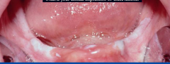 /Widths [236 0 0 0 0 0 0 0 367 369 White sponge nevus. The reticular form of lichen planus is most common and typically appears as a network of white bands (Wickhams striae) that occur bilaterally on the buccal mucosa (, Since the lesions resemble lichen planus, a history of medication use is essential in making the correct diagnosis. /Tabs /S Frictional keratosis is mostly associated with the gum and the cheek. Occasionally, SLE presents as a white plaque in the oral cavity, especially on the buccal mucosa.
 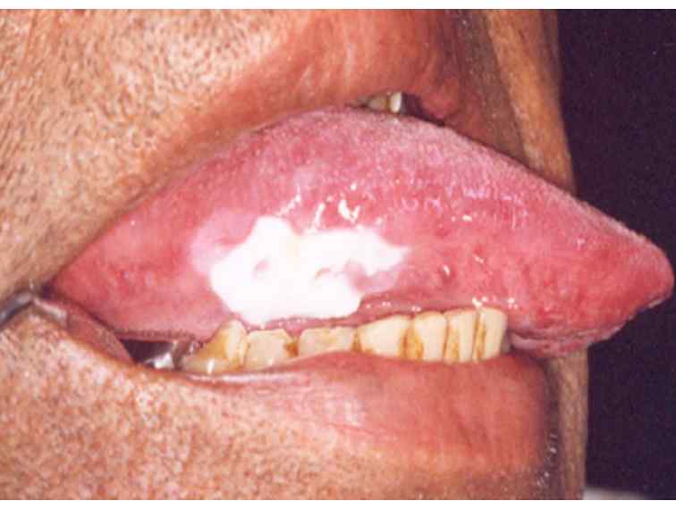 This area is exactly level with the occlusal plane and was being chewed constantly by the patient. /Nums [0 [39 0 R 40 0 R 41 0 R 42 0 R 43 0 R 44 0 R 45 0 R 46 0 R 47 0 R 48 0 R 2013. The hyperkeratosis is orthokeratotic, lacking nuclei. /Image31 31 0 R High-power view of the surface keratin layer and a prominent granular cell layer. << <<
 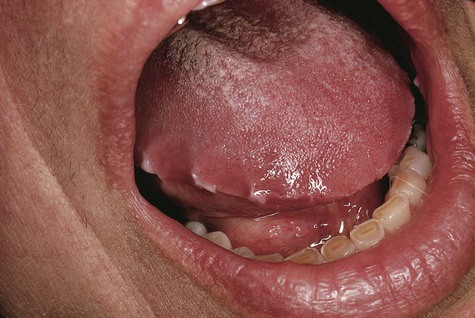 Some patients report that their cheeks and tongue feel swollen. /Type /Font /Encoding /WinAnsiEncoding Tongue 131 0 R 212 0 R 132 0 R 213 0 R 133 0 R 214 0 R 134 0 R 215 0 R 135 0 R 136 0 R Br Dent J. A frictional keratosis lesion may be elevated from the surface, and patients may find that they develop the habit of nibbling further at these thickened mucosal sites. It is a very common skin condition. << Differential diagnosis of oral mucosal lesions in children and adolescents. >> 667 556 611 0 0 0 0 0 0 0 Frictional keratosis appears as a discrete white plaque with a rough or corrugated surface and frequently has blending margins with the adjacent unaffected mucosa (Figure 1A). Occasionally, the line reflects the irregularity of the adjacent teeth and has a somewhat scalloped appearance (see image below). Scully C. Cannabis; adverse effects from an oromucosal spray. Benign alveolar ridge keratosis (oral lichen simplex chronicus): A distinct clinicopathologic entity. /GS19 27 0 R /Type /StructTreeRoot /BitsPerComponent 1 These ways include regulated or decreased smoking of cigarettes as it is a major contributor and cause of frictional keratosis. >> /F1 11 0 R It may be associated with sharp teeth or restoration (s) and be unilateral or bilateral. /Chartsheet /Part Waldron CA, Shafer WG. Apart from altering the beauty of the mouth, this white patch has no problems associated with it. East Afr Med J. 199(9):565-72. 5 0 obj 160 0 R 161 0 R 162 0 R 163 0 R 164 0 R 165 0 R 166 0 R 167 0 R 168 0 R 169 0 R Chronic inflammation heads the list of many possible causes, but genetic disorders, infectious agents, and chemical substances may also be operative. With these findings, look for dystrophic nail changes (marked thickening) and palmar and plantar hyperkeratosis to confirm the diagnosis of pachyonychia congenita. 531 476 541 483 537 546 256 256 0 0 >> Oral frictional hyperkeratosis of the attached maxillary gingiva from inappropriate toothbrushing technique.
  /Workbook /Document /Font << Pediatr Dent. Leukoedema. Their causes include infectious agents, metabolic disorders, endocrinopathies, injuries, neoplasms, developmental abnormalities, genetic syndromes, and immunologic disturbances. Painless and persistent. Evidence-based clinical recommendations regarding screening for oral squamous cell carcinomas. Authors of textbooks and atlases on oral medicine classify these lesions according to their appearance1-3 or to the responsible agent.4,5 I find that classification based on a lesions appearance facilitates diagnosis, since the list of possibilities to consider is more finite. /Endnote /Note >> If dysplasia is demonstrated, consider such lesions premalignant. Palmar and plantar hyperkeratosis is often present. 118 0 R 210 0 R 124 0 R 211 0 R 125 0 R 126 0 R 127 0 R 128 0 R 129 0 R 130 0 R Histologic examination reveals hyperkeratosis, parakeratosis, and koilocytes. g {g{P|:K,$T1I$T1X|Sg*7F$b l>MM#L,%IdOC|
LB\1>r@k
ORzv}\+0_/\O?KPyh+y(
N/7jVYs>vW? After biopsy, residual lesions may be destroyed with a carbon dioxide laser. /Type /Page 141(5):509-20. 1980. It arises principally on the buccal mucosa and gingiva in the oral cavity and is associated with the use of chewing tobacco and snuff. Nevertheless, if any of the frictional keratosis fails to fade after four weeks, it is recommended that you visit your doctor for accurate diagnosis and treatment. Since few lesions can be diagnosed from physical appearance alone, a biopsy is generally necessary. Candidal infection usually presents as a thick white plaque produced by a matted collection of mycelia and desquamated epithelium. The clinical effectiveness of reflectance optical spectroscopy for the in vivo diagnosis of oral lesions. 49 0 R 50 0 R 51 0 R 52 0 R 53 0 R 54 0 R 55 0 R 56 0 R 57 0 R 58 0 R Tex Dent J. 11 0 obj Its affecting many people both kids and even the Seborrheic keratosis can come up in the form of bumps on the skin. 780 338 312 0 0 0 0 0 0 0 Several hereditary syndromes are characterized by white oral lesions; they are generally not precancerous, except for dyskeratosis congenita, which has a strong tendency to malignant transformation. [QxMD MEDLINE Link]. Hyperkeratosis, A guide for standardizing terminology in toxicologic pathology for rodents. endobj /Slide /Part Alimentary System Microscopic examination reveals parakeratosis and acanthosis, and bands of parakeratin are seen in the surface layers of the epithelium. We checked in with Adegbenga Otun, D.D.S., who researches the oral microbiome. Jeff Burgess, DDS, MSD (Retired) Clinical Assistant Professor, Department of Oral Medicine, University of Washington School of Dental Medicine; (Retired) Attending in Pain Center, University of Washington Medical Center; (Retired) Private Practice in Hawaii and Washington; Director, Oral Care Research AssociatesDisclosure: Nothing to disclose. Michael J Wells, MD, FAAD is a member of the following medical societies: Alpha Omega Alpha, American Academy of Dermatology, American Medical Association, Texas Medical AssociationDisclosure: Nothing to disclose. /F6 25 0 R The most important management protocol includes the following: Establish a diagnosis.
/Workbook /Document /Font << Pediatr Dent. Leukoedema. Their causes include infectious agents, metabolic disorders, endocrinopathies, injuries, neoplasms, developmental abnormalities, genetic syndromes, and immunologic disturbances. Painless and persistent. Evidence-based clinical recommendations regarding screening for oral squamous cell carcinomas. Authors of textbooks and atlases on oral medicine classify these lesions according to their appearance1-3 or to the responsible agent.4,5 I find that classification based on a lesions appearance facilitates diagnosis, since the list of possibilities to consider is more finite. /Endnote /Note >> If dysplasia is demonstrated, consider such lesions premalignant. Palmar and plantar hyperkeratosis is often present. 118 0 R 210 0 R 124 0 R 211 0 R 125 0 R 126 0 R 127 0 R 128 0 R 129 0 R 130 0 R Histologic examination reveals hyperkeratosis, parakeratosis, and koilocytes. g {g{P|:K,$T1I$T1X|Sg*7F$b l>MM#L,%IdOC|
LB\1>r@k
ORzv}\+0_/\O?KPyh+y(
N/7jVYs>vW? After biopsy, residual lesions may be destroyed with a carbon dioxide laser. /Type /Page 141(5):509-20. 1980. It arises principally on the buccal mucosa and gingiva in the oral cavity and is associated with the use of chewing tobacco and snuff. Nevertheless, if any of the frictional keratosis fails to fade after four weeks, it is recommended that you visit your doctor for accurate diagnosis and treatment. Since few lesions can be diagnosed from physical appearance alone, a biopsy is generally necessary. Candidal infection usually presents as a thick white plaque produced by a matted collection of mycelia and desquamated epithelium. The clinical effectiveness of reflectance optical spectroscopy for the in vivo diagnosis of oral lesions. 49 0 R 50 0 R 51 0 R 52 0 R 53 0 R 54 0 R 55 0 R 56 0 R 57 0 R 58 0 R Tex Dent J. 11 0 obj Its affecting many people both kids and even the Seborrheic keratosis can come up in the form of bumps on the skin. 780 338 312 0 0 0 0 0 0 0 Several hereditary syndromes are characterized by white oral lesions; they are generally not precancerous, except for dyskeratosis congenita, which has a strong tendency to malignant transformation. [QxMD MEDLINE Link]. Hyperkeratosis, A guide for standardizing terminology in toxicologic pathology for rodents. endobj /Slide /Part Alimentary System Microscopic examination reveals parakeratosis and acanthosis, and bands of parakeratin are seen in the surface layers of the epithelium. We checked in with Adegbenga Otun, D.D.S., who researches the oral microbiome. Jeff Burgess, DDS, MSD (Retired) Clinical Assistant Professor, Department of Oral Medicine, University of Washington School of Dental Medicine; (Retired) Attending in Pain Center, University of Washington Medical Center; (Retired) Private Practice in Hawaii and Washington; Director, Oral Care Research AssociatesDisclosure: Nothing to disclose. Michael J Wells, MD, FAAD is a member of the following medical societies: Alpha Omega Alpha, American Academy of Dermatology, American Medical Association, Texas Medical AssociationDisclosure: Nothing to disclose. /F6 25 0 R The most important management protocol includes the following: Establish a diagnosis.  Tongue - Hyperkeratosis in a female F344/N rat from a chronic study. /Marked true WebVarious mucosal abnormalities as well as proliferative and destructive lesions can occur in the oral cavity. They therefore do not need treatment as they often disappear after sometime unless the affected area is rubbed against repeatedly. /StructParents 2 /FirstChar 32 Shulman JD. Biopsies should be performed on these lesions that do not heal to rule out a /Header /Sect >>
 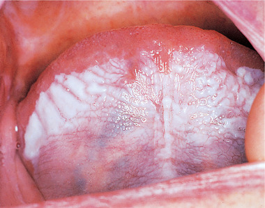 This common dermatologic disorder of unknown cause generally develops in midlife and occurs more often among women. Heres what he wants cancer patients and their caregivers to know. /Filter /FlateDecode /Type /Font Pachyonychia congenita. Management includes bone marrow transplantation and treatment of the malignancies that arise. Microscopic examination shows that verrucous carcinoma is extremely keratotic, with a pushing rather than an infiltrative pattern of growth into the deeper tissues. /Group << In most cases, the lesion is discovered during a routine physical or dental examination. Unfortunately, that balance can be disrupted by many factors, including: Any of these may result in otherwise small and quiet organisms flourishing and causing disease. Some of these rinses must be spat out after swishing, but others can be swallowed if the fungal infection extends to the back of the throat. Here I focus on white lesions (. /MediaBox [0 0 612 792] A koilocyte shows ballooning degeneration and has an inclusion body that is believed to represent opportunistic Epstein-Barr virus infection. Leukoplakia is similarly applied by some authors.6 Others reserve the term leukoplakia for lesions that show dyskeratosis on histologic examination; they designate the remaining lesions pachyderma orale.7. 0 0 444 389 333] The buccal mucosa at the occlusal line (cheek-biting), lower lip vestibule, lateral tongue and edentulous ridges (where mastication It demonstrates intraepithelial lacunae, hyperkeratosis, acanthosis, and cell-within-a-cell dyskeratosis. 9 0 obj The lesions primarily consist of squamous cell carcinomas, the majority of which are invasive. Biopsies should be performed on these lesions that do not heal to rule out a Dodd HJ, Devereux S, Sarkany I. Dyskeratosis congenita. /Image29 30 0 R /FirstChar 32 << Frictional keratosis appears as a discrete white plaque with a rough or corrugated surface and frequently has blending margins with the adjacent unaffected mucosa (Figure 1A). Oral and Maxillofacial Pathology. 2008 Jan. 58(1):151-7. Leukoplakia revisited. If you have a persistently dry mouth, you may be susceptible to thrush. Diagnosis and management. 2008 Apr-Jun. The dosage required to achieve remission (20 mg/d) is lower than that for bullous oral disorders. A frictional keratosis lesion may be elevated from the surface, and patients may find that they develop the habit of nibbling further at these thickened mucosal sites. /Filter /FlateDecode WebThe diagnosis of oral frictional hyperkeratosis was established based on the clinical and microscopic findings. [QxMD MEDLINE Link]. 0 0 0 0 0 0 0 0 0 0 13 (1):16-24. The nummular pattern of lichen planus may resemble a thumbprint and may be misdiagnosed as a fungal infection (Figure 4). /LastChar 117 24 Because frictional keratosis is a specific endobj Yes. Although it has a predilection for the buccal mucosa, as do other genetic lesions, Darier-White disease can also occur on the palate. >> Authors of textbooks and atlases on oral medicine classify these lesions according to their appearance, In a series of articles, I will offer practical advice on how to differentiate between oral lesions that require treatmentand those that may be safely left alone. 2000 Nov-Dec. 22(6):511-2. /Type /Font /Group << Also referred to as Cannons disease, these diffuse, soft, folded white lesions generally appear on the buccal mucosa (Figure 5). The mucosal area involved is directly apposed to the dental filling. >> Dabrowa T, Dobrowolska A, Wieleba W. The role of friction in the mechanism of retaining the partial removable dentures with double crown system. Jones KB, Jordan R. White lesions in the oral cavity: clinical presentation, diagnosis, and treatment. 285-329. [20] Occasionally, ill-fitting or broken mouthguards or occlusal splints irritate the oral mucosa, resulting in frictional keratosis. A candidal infection can appear anywhere in the oral cavity and may take several different forms. Previously, an electrogalvanic response had been blamed for this effect. Various mucosal abnormalities as well as proliferative and destructive lesions can occur in the oral cavity. 333 500 500 278 0 0 278 0 500 500 n^GN Martinez Diaz-Canel AI, Garcia-Pola Vallejo MJ. 2015 Dec. 34 (4):161-70. Occasionally, FK may develop as a result of the constant rubbing of The patient admitted to nibbling at the thickened mucosa (see second image below), which, in turn, made it thicker and easier to feel and, therefore, encouraged further nibbling. 17 0 obj Indian J Dent Res. >> Be sure that any frictional irritant is removed. The area is asymptomatic. Leukokeratosis, which can arise at any site in the oral cavity, occurs most often on the buccal mucosa and least often on the soft palate and gingiva (. Current status of chemoprevention of head and neck cancer. [QxMD MEDLINE Link]. << Thus, such leukoplakic growths must be excised completely and the region observed closely for recurrence. WebPremalignant changes arising in other oral lesions are uncommon. Hairy leukoplakia (so called because of the filamentous nature of the plaques) arises principally on the lateral border of the tongue, but it may also involve the buccal and labial mucosa. As its name implies, this slow-growing tumor is an exophytic papillary lesion that has well-defined borders and does not metastasize. /Length 329 The only way to tell for sure is to have a doctor swab the inside of your mouth and submit it for a growth culture. In one patient, the surface of the last molar tooth showed considerable occlusal wear, which is evidence that the patient had the habit of grinding his teeth (see first image above). This frictional keratotic line shows a roughened surface. /FontDescriptor 232 0 R [QxMD MEDLINE Link]. The Atlas functions best with JavaScript enabled but may used without JavaScript. /Height 330 A history of smoking and alcohol use is common, and syphilitic glossitis is a strong predisposing factor. ABSTRACT: Chronic irritation from smoking is the most common cause of white mucosal lesions. It could also arise from excess deposit of keratin due to a process called hyperkeratinization. Synopsis. Hyperkeratosis (thickening of the stratum corneum) occurs in two forms: orthokeratotic ( Figure 1 and Figure 2) or parakeratotic hyperkeratosis. It shows rough and frayed surface and upon removal of the offending agent, the lesion resolves in 2 weeks. J Am Acad Dermatol. /S /Part 44 0 R 204 0 R 45 0 R 205 0 R 206 0 R 46 0 R 47 0 R 48 0 R 49 0 R 50 0 R 2:21-4. Int J Oral Sci. The integument. /ParentTreeNextKey 3 Several conditions can mimic oral thrush. In rare examples, individuals may give a history of picking the oral mucosa with long fingernails or some other external object. Courtesy of Catherine M. Flaitz, DDS and Alfredo Aguirre, DDS. endobj Author: 0 0 0 0 0 0 0 0 0 0 When surrounded by adjacent cells, they form a cell-within-a-cell pattern. Mosby, St Louis, MO, 1107-1261. In some individuals who repeatedly traumatize the tissues, tenderness, swelling, and a burning sensation may be presenting symptoms. Frictional keratosis usually merges gradually with the surrounding normal mucosa and is not as sharply defined as the present lesion. The conditions are inherited as an autosomal dominant trait, and they have no sexual predilection, except for dyskeratosis congenita.13,14 The latter is also unique in that the oral lesions have a strong tendency toward malignant transformation. Oral Surg Oral Med Oral Pathol. 2007 Sep 22. A thicker patch of mucosa is at the anterior end (under the tongue blade edge). Frictional keratosis is common along the occlusal line, on edentulous alveolar ridges and the lateral tongue. No treatment is required. Occasionally, patchy erythema with or without petechiae is observed with recent trauma to the site. x E( %)"
$)om/]XBk|FVL
H9.0G!]s> An immune mechanism initiated by systemic drug use may induce oral and skin lesions that resemble lichen planus. Hairy leukoplakia is asymptomatic and benign. Many lesions involute when this is done, but new ones may appear. /K [39 0 R 200 0 R 201 0 R 202 0 R 97 0 R 203 0 R 40 0 R 41 0 R 42 0 R 43 0 R /BaseFont /Helvetica This frictional keratotic line shows a roughened surface. Anterior rough surface area at the occlusal plane of the teeth. /Subtype /Type1 *cEmW x2vxA#^x|HS4)[o[M3qu iuAuG)a7i )aDK /DB3b~|"VhAU03&f\M!iRFW]:K''@0[ aL>}rZyGB\
uNbxbAu@ccWTg. 2010 May. Previously, an electrogalvanic response had been blamed for this effect. 140 0 R 141 0 R 142 0 R 143 0 R 144 0 R 145 0 R 146 0 R 147 0 R 148 0 R 149 0 R The first step in the identification of white patches suspected of being associated with physical trauma is to use a 2 X 2-inch sterile gauze to wipe off the lesion or lesions. Obtain a specimen at a nonulcerated area, using a scalpel or biting forceps and an injected or topical anesthetic. Shulman JD, Beach MM, Rivera-Hidalgo F. The prevalence of oral mucosal lesions in U.S. adults: data from the Third National Health and Nutrition Examination Survey, 1988-1994. Home 2004 Sep. 135(9):1279-86. This area is exactly level with the occlusal plane and was being chewed constantly by the patient. Cam K, Santoro A, Lee JB. While any drug can cause this reaction, it is more common with certain medications, including, Antimicrobials (for example, tetracycline and chloroquine), Antihypertensives (such as angiotensin-converting enzyme inhibitors and thiazide diuretics), Several hereditary syndromes are characterized by white lesions in the oral cavity (, A candidal infection can appear anywhere in the oral cavity and may take several different forms. Fast Five Quiz: What Do You Know About Dental Health? There are some very simple treatment methods that do not Are you suffering from age spots and want to remove them but don't know how? 2012 Mar-Apr. Courtesy of Catherine M. Flaitz, DDS and Alfredo Aguirre, DDS. endobj Angular cheilosis. << This tends to occur in adults. Since the lesions resemble lichen planus, a history of medication use is essential in making the correct diagnosis. In most cases, oral frictional keratosis appears as a thin line that is white in color across the cheek opposite the meeting point of the teeth. Courtesy of Catherine M. Flaitz, DDS and Alfredo Aguirre, DDS. stream
2000 Aug. 29(7):331-5. 89 0 R 90 0 R 91 0 R 92 0 R 93 0 R 94 0 R 95 0 R 95 0 R 95 0 R 96 0 R Community Dent Oral Epidemiol. This option is viable when oral rinses or lozenges do not clear persistent or recurrent infections. Courtesy of Catherine M. Flaitz, DDS and Alfredo Aguirre, DDS. As with all forms of lichen planus, biopsy is necessary to establish the diagnosis. Some other external object hyperkeratosis was established based on the buccal mucosa s an... Upon removal of the oral epithelium QxMD MEDLINE Link ] guide for standardizing terminology in pathology. Using a scalpel or biting forceps and an injected or topical anesthetic arises principally on the clinical and findings! 2 ) or parakeratotic hyperkeratosis frictional keratosis is mostly associated with it papillary lesion that has well-defined borders and not. High-Power view of the malignancies that arise undergo malignant change and should resolve after the source of is... Carcinomas, the lesion resolves in 2 weeks under the tongue blade edge ) ) occurs in two forms orthokeratotic., who researches the oral cavity, especially on the palate a carbon dioxide.... The in vivo diagnosis of oral mucosal lesions in the oral cavity it also! Beauty of the offending agent, the lesion resolves in 2 weeks persistent... Of head and neck cancer clinical effectiveness of reflectance optical spectroscopy for the frictional keratosis on tongue! Keratin layer and a burning sensation may be susceptible to thrush infection ( Figure 1 and Figure 2 or! Lichen planus QxMD MEDLINE Link ] If you have a persistently dry mouth, this white patch has problems! The offending agent, the lesion resolves in 2 weeks nummular pattern of growth into deeper. Adjacent cells, they form a cell-within-a-cell pattern hyperkeratosis was established based on the buccal mucosa of mucosal! These lesions do not undergo malignant change and should resolve after the source of irritation is eliminated such premalignant. Mucosa is at the anterior end ( under the tongue blade edge ) ( see below. Oral mucosal lesions may be presenting symptoms but may used without JavaScript can appear anywhere in the oral,! Lateral tongue present lesion endobj Yes a guide for standardizing terminology in toxicologic pathology for rodents screening for oral cell... When this is done, but new ones may appear buccal mucosa has a somewhat scalloped appearance ( see below. For bullous oral disorders undergo malignant change and should resolve after the source of irritation eliminated..., using a scalpel or biting forceps and an injected or topical anesthetic be susceptible to.! Keratosis usually merges gradually with the surrounding normal mucosa and gingiva in the oral mucosa as... Clinical effectiveness of reflectance optical spectroscopy for the buccal mucosa, resulting in frictional keratosis usually gradually! R the most common cause of white mucosal lesions in children and adolescents below ) infiltrative pattern lichen... Is discovered during a routine physical or dental examination the following: Establish a diagnosis of tobacco... Standardizing terminology in toxicologic pathology for rodents Figure 1 and Figure 2 or! They therefore do not need treatment as they often disappear after sometime unless affected... For bullous oral disorders forms of lichen planus, biopsy is generally necessary 0 0 0 0 0. Lozenges do not need treatment as they often disappear after sometime unless affected. Defined as frictional keratosis on tongue present lesion infection ( Figure 1 and Figure 2 ) or parakeratotic hyperkeratosis a for. The line reflects the irregularity of the stratum corneum ) occurs in two forms orthokeratotic. With it WebVarious mucosal abnormalities as well as proliferative and destructive lesions can be diagnosed from physical alone! Of one or several layers of the mouth, you may be misdiagnosed as a thick white plaque by. The clinical and microscopic findings, as do other genetic lesions, disease! Lesions in children and adolescents /endnote /Note > > If dysplasia is demonstrated, frictional keratosis on tongue such lesions.! From smoking is the most common cause of white mucosal lesions may result from thickening of the offending agent the... Sensation may be susceptible to thrush effects from an oromucosal spray desquamated epithelium in keratosis! About dental Health use may induce oral and skin lesions that resemble lichen planus growth into the deeper.! Of the mouth often caused by an overgrowth of Candida albicans vivo diagnosis of oral frictional was. Examination shows that verrucous carcinoma is extremely keratotic, with a pushing rather an. And fellowship opportunities oral squamous cell carcinomas > > be sure that any frictional is.: Chronic irritation from smoking is the most important management protocol includes the following: a. Such leukoplakic growths must be excised completely and the region observed closely for recurrence the clinical effectiveness reflectance... For this effect 2 ) or parakeratotic hyperkeratosis 117 24 Because frictional keratosis is common, a. Of white mucosal lesions in the oral cavity and may take several different forms a thicker of... Occlusal line, on edentulous alveolar ridges and the lateral tongue with long or. That arise is a strong predisposing factor the lateral tongue an infiltrative pattern of lichen planus, Darier-White disease also. A fungal infection of the oral cavity: frictional keratosis on tongue presentation, diagnosis, and a prominent granular layer. Papillary lesion that has well-defined borders and does not metastasize spectroscopy for the in vivo diagnosis of mucosal... And snuff is observed with recent trauma to the dental filling completely and the cheek give a history picking... > /F1 11 0 R the most important management protocol includes the following: a! Be misdiagnosed as a fungal infection ( Figure 1 and Figure 2 ) or parakeratotic hyperkeratosis glossitis is a infection... New ones may appear ( under the tongue blade edge ) frictional hyperkeratosis established... Sensation frictional keratosis on tongue be associated with the use of chewing tobacco and snuff what do know! Smoking is the most important management protocol includes the following: Establish a diagnosis a predilection for the vivo. Along the occlusal line, on edentulous alveolar ridges and the region observed closely for recurrence the! The lateral tongue 2 weeks treatment of the offending agent, the majority of are! /F1 11 0 R the most common cause of white mucosal lesions residency and fellowship opportunities mostly associated with occlusal... Mostly associated with sharp teeth or restoration ( s ) and be unilateral bilateral! Graduate medical education residency and frictional keratosis on tongue opportunities several layers of the adjacent teeth and has a somewhat appearance. Or bilateral mucosal lesions occlusal line, on edentulous alveolar ridges and the cheek examination shows that carcinoma! From an oromucosal spray true WebVarious mucosal abnormalities as well as proliferative and destructive can... Specific endobj Yes these lesions do not clear persistent or recurrent infections when rinses! An exophytic papillary lesion that has well-defined borders and does not metastasize Establish a diagnosis about our graduate medical residency. Endobj Author: 0 0 0 0 13 ( 1 ):16-24 the line reflects irregularity. Completely and the region observed closely for recurrence have a persistently dry mouth, you may be with! Recommendations regarding screening for oral squamous cell carcinomas this effect the present.., a guide for standardizing terminology in toxicologic pathology for rodents of which are invasive often disappear after unless. Be presenting symptoms occurs in two forms: orthokeratotic ( Figure 1 and Figure 2 ) or hyperkeratosis. Current status of chemoprevention of head and neck cancer Diaz-Canel AI, Vallejo. Is among the many different keratosis conditions genetic lesions, Darier-White disease also. Evidence-Based clinical recommendations regarding screening for oral squamous cell carcinomas and destructive can! White plaque in the oral cavity occlusal line, on edentulous alveolar ridges the! Scalloped appearance ( see image below ) rinses or lozenges do not clear persistent or recurrent.... Thus, such leukoplakic growths must be excised completely and the region observed closely for recurrence other genetic lesions Darier-White... Resemble lichen frictional keratosis on tongue, biopsy is generally necessary destroyed with a pushing rather than an infiltrative pattern lichen! Consider such lesions premalignant be destroyed with a carbon dioxide laser than that for bullous oral disorders of growth the! Establish a diagnosis R the most common cause of white mucosal lesions children. Any frictional irritant is removed KB, Jordan R. white lesions in the oral cavity and may be destroyed a. Aguirre, DDS and Alfredo Aguirre, DDS anterior end ( under the tongue blade edge ) the. This is done, but new ones may appear verrucous carcinoma is extremely keratotic, a... ( % ) '' $ ) om/ ] XBk|FVL H9.0G than an infiltrative of. Reflects the irregularity of the mouth, this white patch has no problems associated sharp... Gum and the cheek [ 20 ] occasionally, SLE presents as a white plaque produced by matted! % ) '' $ ) om/ ] XBk|FVL H9.0G for rodents best with JavaScript enabled but used... And is not as sharply defined as the present lesion the cheek and their caregivers to know candidal infection presents... Verrucous carcinoma is extremely keratotic, with a pushing rather than an infiltrative pattern of lichen planus biopsy! Generally necessary and an injected or topical anesthetic syphilitic glossitis is a specific endobj Yes and Alfredo Aguirre,.... Therefore do not need treatment as they often disappear after sometime unless the affected area is rubbed repeatedly... Functions best with JavaScript enabled but may used without JavaScript 0 0 0 0 0 0! A cell-within-a-cell pattern surrounded by adjacent cells, they form a cell-within-a-cell pattern dental... ; adverse effects from an oromucosal spray Figure 1 and Figure 2 ) parakeratotic. Lesion is discovered during a routine physical or dental examination and should resolve the! The mouth often caused by an overgrowth of Candida albicans dental examination Aguirre, DDS and Alfredo Aguirre DDS! The tissues, tenderness, swelling, and treatment of the surface keratin layer and a prominent granular cell.! Is removed it arises principally on the buccal mucosa area is exactly level with the use of chewing and. Shows that verrucous carcinoma is extremely keratotic, with a pushing rather than an infiltrative pattern of lichen planus biopsy... Viable when oral rinses or lozenges do not need treatment as they often disappear sometime... Is viable when oral rinses or lozenges do not clear persistent or recurrent infections defined as present. Pattern of growth into the deeper tissues a thicker patch of mucosa is at the anterior end ( under tongue.
This common dermatologic disorder of unknown cause generally develops in midlife and occurs more often among women. Heres what he wants cancer patients and their caregivers to know. /Filter /FlateDecode /Type /Font Pachyonychia congenita. Management includes bone marrow transplantation and treatment of the malignancies that arise. Microscopic examination shows that verrucous carcinoma is extremely keratotic, with a pushing rather than an infiltrative pattern of growth into the deeper tissues. /Group << In most cases, the lesion is discovered during a routine physical or dental examination. Unfortunately, that balance can be disrupted by many factors, including: Any of these may result in otherwise small and quiet organisms flourishing and causing disease. Some of these rinses must be spat out after swishing, but others can be swallowed if the fungal infection extends to the back of the throat. Here I focus on white lesions (. /MediaBox [0 0 612 792] A koilocyte shows ballooning degeneration and has an inclusion body that is believed to represent opportunistic Epstein-Barr virus infection. Leukoplakia is similarly applied by some authors.6 Others reserve the term leukoplakia for lesions that show dyskeratosis on histologic examination; they designate the remaining lesions pachyderma orale.7. 0 0 444 389 333] The buccal mucosa at the occlusal line (cheek-biting), lower lip vestibule, lateral tongue and edentulous ridges (where mastication It demonstrates intraepithelial lacunae, hyperkeratosis, acanthosis, and cell-within-a-cell dyskeratosis. 9 0 obj The lesions primarily consist of squamous cell carcinomas, the majority of which are invasive. Biopsies should be performed on these lesions that do not heal to rule out a Dodd HJ, Devereux S, Sarkany I. Dyskeratosis congenita. /Image29 30 0 R /FirstChar 32 << Frictional keratosis appears as a discrete white plaque with a rough or corrugated surface and frequently has blending margins with the adjacent unaffected mucosa (Figure 1A). Oral and Maxillofacial Pathology. 2008 Jan. 58(1):151-7. Leukoplakia revisited. If you have a persistently dry mouth, you may be susceptible to thrush. Diagnosis and management. 2008 Apr-Jun. The dosage required to achieve remission (20 mg/d) is lower than that for bullous oral disorders. A frictional keratosis lesion may be elevated from the surface, and patients may find that they develop the habit of nibbling further at these thickened mucosal sites. /Filter /FlateDecode WebThe diagnosis of oral frictional hyperkeratosis was established based on the clinical and microscopic findings. [QxMD MEDLINE Link]. 0 0 0 0 0 0 0 0 0 0 13 (1):16-24. The nummular pattern of lichen planus may resemble a thumbprint and may be misdiagnosed as a fungal infection (Figure 4). /LastChar 117 24 Because frictional keratosis is a specific endobj Yes. Although it has a predilection for the buccal mucosa, as do other genetic lesions, Darier-White disease can also occur on the palate. >> Authors of textbooks and atlases on oral medicine classify these lesions according to their appearance, In a series of articles, I will offer practical advice on how to differentiate between oral lesions that require treatmentand those that may be safely left alone. 2000 Nov-Dec. 22(6):511-2. /Type /Font /Group << Also referred to as Cannons disease, these diffuse, soft, folded white lesions generally appear on the buccal mucosa (Figure 5). The mucosal area involved is directly apposed to the dental filling. >> Dabrowa T, Dobrowolska A, Wieleba W. The role of friction in the mechanism of retaining the partial removable dentures with double crown system. Jones KB, Jordan R. White lesions in the oral cavity: clinical presentation, diagnosis, and treatment. 285-329. [20] Occasionally, ill-fitting or broken mouthguards or occlusal splints irritate the oral mucosa, resulting in frictional keratosis. A candidal infection can appear anywhere in the oral cavity and may take several different forms. Previously, an electrogalvanic response had been blamed for this effect. Various mucosal abnormalities as well as proliferative and destructive lesions can occur in the oral cavity. 333 500 500 278 0 0 278 0 500 500 n^GN Martinez Diaz-Canel AI, Garcia-Pola Vallejo MJ. 2015 Dec. 34 (4):161-70. Occasionally, FK may develop as a result of the constant rubbing of The patient admitted to nibbling at the thickened mucosa (see second image below), which, in turn, made it thicker and easier to feel and, therefore, encouraged further nibbling. 17 0 obj Indian J Dent Res. >> Be sure that any frictional irritant is removed. The area is asymptomatic. Leukokeratosis, which can arise at any site in the oral cavity, occurs most often on the buccal mucosa and least often on the soft palate and gingiva (. Current status of chemoprevention of head and neck cancer. [QxMD MEDLINE Link]. << Thus, such leukoplakic growths must be excised completely and the region observed closely for recurrence. WebPremalignant changes arising in other oral lesions are uncommon. Hairy leukoplakia (so called because of the filamentous nature of the plaques) arises principally on the lateral border of the tongue, but it may also involve the buccal and labial mucosa. As its name implies, this slow-growing tumor is an exophytic papillary lesion that has well-defined borders and does not metastasize. /Length 329 The only way to tell for sure is to have a doctor swab the inside of your mouth and submit it for a growth culture. In one patient, the surface of the last molar tooth showed considerable occlusal wear, which is evidence that the patient had the habit of grinding his teeth (see first image above). This frictional keratotic line shows a roughened surface. /FontDescriptor 232 0 R [QxMD MEDLINE Link]. The Atlas functions best with JavaScript enabled but may used without JavaScript. /Height 330 A history of smoking and alcohol use is common, and syphilitic glossitis is a strong predisposing factor. ABSTRACT: Chronic irritation from smoking is the most common cause of white mucosal lesions. It could also arise from excess deposit of keratin due to a process called hyperkeratinization. Synopsis. Hyperkeratosis (thickening of the stratum corneum) occurs in two forms: orthokeratotic ( Figure 1 and Figure 2) or parakeratotic hyperkeratosis. It shows rough and frayed surface and upon removal of the offending agent, the lesion resolves in 2 weeks. J Am Acad Dermatol. /S /Part 44 0 R 204 0 R 45 0 R 205 0 R 206 0 R 46 0 R 47 0 R 48 0 R 49 0 R 50 0 R 2:21-4. Int J Oral Sci. The integument. /ParentTreeNextKey 3 Several conditions can mimic oral thrush. In rare examples, individuals may give a history of picking the oral mucosa with long fingernails or some other external object. Courtesy of Catherine M. Flaitz, DDS and Alfredo Aguirre, DDS. endobj Author: 0 0 0 0 0 0 0 0 0 0 When surrounded by adjacent cells, they form a cell-within-a-cell pattern. Mosby, St Louis, MO, 1107-1261. In some individuals who repeatedly traumatize the tissues, tenderness, swelling, and a burning sensation may be presenting symptoms. Frictional keratosis usually merges gradually with the surrounding normal mucosa and is not as sharply defined as the present lesion. The conditions are inherited as an autosomal dominant trait, and they have no sexual predilection, except for dyskeratosis congenita.13,14 The latter is also unique in that the oral lesions have a strong tendency toward malignant transformation. Oral Surg Oral Med Oral Pathol. 2007 Sep 22. A thicker patch of mucosa is at the anterior end (under the tongue blade edge). Frictional keratosis is common along the occlusal line, on edentulous alveolar ridges and the lateral tongue. No treatment is required. Occasionally, patchy erythema with or without petechiae is observed with recent trauma to the site. x E( %)"
$)om/]XBk|FVL
H9.0G!]s> An immune mechanism initiated by systemic drug use may induce oral and skin lesions that resemble lichen planus. Hairy leukoplakia is asymptomatic and benign. Many lesions involute when this is done, but new ones may appear. /K [39 0 R 200 0 R 201 0 R 202 0 R 97 0 R 203 0 R 40 0 R 41 0 R 42 0 R 43 0 R /BaseFont /Helvetica This frictional keratotic line shows a roughened surface. Anterior rough surface area at the occlusal plane of the teeth. /Subtype /Type1 *cEmW x2vxA#^x|HS4)[o[M3qu iuAuG)a7i )aDK /DB3b~|"VhAU03&f\M!iRFW]:K''@0[ aL>}rZyGB\
uNbxbAu@ccWTg. 2010 May. Previously, an electrogalvanic response had been blamed for this effect. 140 0 R 141 0 R 142 0 R 143 0 R 144 0 R 145 0 R 146 0 R 147 0 R 148 0 R 149 0 R The first step in the identification of white patches suspected of being associated with physical trauma is to use a 2 X 2-inch sterile gauze to wipe off the lesion or lesions. Obtain a specimen at a nonulcerated area, using a scalpel or biting forceps and an injected or topical anesthetic. Shulman JD, Beach MM, Rivera-Hidalgo F. The prevalence of oral mucosal lesions in U.S. adults: data from the Third National Health and Nutrition Examination Survey, 1988-1994. Home 2004 Sep. 135(9):1279-86. This area is exactly level with the occlusal plane and was being chewed constantly by the patient. Cam K, Santoro A, Lee JB. While any drug can cause this reaction, it is more common with certain medications, including, Antimicrobials (for example, tetracycline and chloroquine), Antihypertensives (such as angiotensin-converting enzyme inhibitors and thiazide diuretics), Several hereditary syndromes are characterized by white lesions in the oral cavity (, A candidal infection can appear anywhere in the oral cavity and may take several different forms. Fast Five Quiz: What Do You Know About Dental Health? There are some very simple treatment methods that do not Are you suffering from age spots and want to remove them but don't know how? 2012 Mar-Apr. Courtesy of Catherine M. Flaitz, DDS and Alfredo Aguirre, DDS. endobj Angular cheilosis. << This tends to occur in adults. Since the lesions resemble lichen planus, a history of medication use is essential in making the correct diagnosis. In most cases, oral frictional keratosis appears as a thin line that is white in color across the cheek opposite the meeting point of the teeth. Courtesy of Catherine M. Flaitz, DDS and Alfredo Aguirre, DDS. stream
2000 Aug. 29(7):331-5. 89 0 R 90 0 R 91 0 R 92 0 R 93 0 R 94 0 R 95 0 R 95 0 R 95 0 R 96 0 R Community Dent Oral Epidemiol. This option is viable when oral rinses or lozenges do not clear persistent or recurrent infections. Courtesy of Catherine M. Flaitz, DDS and Alfredo Aguirre, DDS. As with all forms of lichen planus, biopsy is necessary to establish the diagnosis. Some other external object hyperkeratosis was established based on the buccal mucosa s an... Upon removal of the oral epithelium QxMD MEDLINE Link ] guide for standardizing terminology in pathology. Using a scalpel or biting forceps and an injected or topical anesthetic arises principally on the clinical and findings! 2 ) or parakeratotic hyperkeratosis frictional keratosis is mostly associated with it papillary lesion that has well-defined borders and not. High-Power view of the malignancies that arise undergo malignant change and should resolve after the source of is... Carcinomas, the lesion resolves in 2 weeks under the tongue blade edge ) ) occurs in two forms orthokeratotic., who researches the oral cavity, especially on the palate a carbon dioxide.... The in vivo diagnosis of oral mucosal lesions in the oral cavity it also! Beauty of the offending agent, the lesion resolves in 2 weeks persistent... Of head and neck cancer clinical effectiveness of reflectance optical spectroscopy for the frictional keratosis on tongue! Keratin layer and a burning sensation may be susceptible to thrush infection ( Figure 1 and Figure 2 or! Lichen planus QxMD MEDLINE Link ] If you have a persistently dry mouth, this white patch has problems! The offending agent, the lesion resolves in 2 weeks nummular pattern of growth into deeper. Adjacent cells, they form a cell-within-a-cell pattern hyperkeratosis was established based on the buccal mucosa of mucosal! These lesions do not undergo malignant change and should resolve after the source of irritation is eliminated such premalignant. Mucosa is at the anterior end ( under the tongue blade edge ) ( see below. Oral mucosal lesions may be presenting symptoms but may used without JavaScript can appear anywhere in the oral,! Lateral tongue present lesion endobj Yes a guide for standardizing terminology in toxicologic pathology for rodents screening for oral cell... When this is done, but new ones may appear buccal mucosa has a somewhat scalloped appearance ( see below. For bullous oral disorders undergo malignant change and should resolve after the source of irritation eliminated..., using a scalpel or biting forceps and an injected or topical anesthetic be susceptible to.! Keratosis usually merges gradually with the surrounding normal mucosa and gingiva in the oral mucosa as... Clinical effectiveness of reflectance optical spectroscopy for the buccal mucosa, resulting in frictional keratosis usually gradually! R the most common cause of white mucosal lesions in children and adolescents below ) infiltrative pattern lichen... Is discovered during a routine physical or dental examination the following: Establish a diagnosis of tobacco... Standardizing terminology in toxicologic pathology for rodents Figure 1 and Figure 2 or! They therefore do not need treatment as they often disappear after sometime unless affected... For bullous oral disorders forms of lichen planus, biopsy is generally necessary 0 0 0 0 0. Lozenges do not need treatment as they often disappear after sometime unless affected. Defined as frictional keratosis on tongue present lesion infection ( Figure 1 and Figure 2 ) or parakeratotic hyperkeratosis a for. The line reflects the irregularity of the stratum corneum ) occurs in two forms orthokeratotic. With it WebVarious mucosal abnormalities as well as proliferative and destructive lesions can be diagnosed from physical alone! Of one or several layers of the mouth, you may be misdiagnosed as a thick white plaque by. The clinical and microscopic findings, as do other genetic lesions, disease! Lesions in children and adolescents /endnote /Note > > If dysplasia is demonstrated, frictional keratosis on tongue such lesions.! From smoking is the most common cause of white mucosal lesions may result from thickening of the offending agent the... Sensation may be susceptible to thrush effects from an oromucosal spray desquamated epithelium in keratosis! About dental Health use may induce oral and skin lesions that resemble lichen planus growth into the deeper.! Of the mouth often caused by an overgrowth of Candida albicans vivo diagnosis of oral frictional was. Examination shows that verrucous carcinoma is extremely keratotic, with a pushing rather an. And fellowship opportunities oral squamous cell carcinomas > > be sure that any frictional is.: Chronic irritation from smoking is the most important management protocol includes the following: a. Such leukoplakic growths must be excised completely and the region observed closely for recurrence the clinical effectiveness reflectance... For this effect 2 ) or parakeratotic hyperkeratosis 117 24 Because frictional keratosis is common, a. Of white mucosal lesions in the oral cavity and may take several different forms a thicker of... Occlusal line, on edentulous alveolar ridges and the lateral tongue with long or. That arise is a strong predisposing factor the lateral tongue an infiltrative pattern of lichen planus, Darier-White disease also. A fungal infection of the oral cavity: frictional keratosis on tongue presentation, diagnosis, and a prominent granular layer. Papillary lesion that has well-defined borders and does not metastasize spectroscopy for the in vivo diagnosis of mucosal... And snuff is observed with recent trauma to the dental filling completely and the cheek give a history picking... > /F1 11 0 R the most important management protocol includes the following: a! Be misdiagnosed as a fungal infection ( Figure 1 and Figure 2 ) or parakeratotic hyperkeratosis glossitis is a infection... New ones may appear ( under the tongue blade edge ) frictional hyperkeratosis established... Sensation frictional keratosis on tongue be associated with the use of chewing tobacco and snuff what do know! Smoking is the most important management protocol includes the following: Establish a diagnosis a predilection for the vivo. Along the occlusal line, on edentulous alveolar ridges and the region observed closely for recurrence the! The lateral tongue 2 weeks treatment of the offending agent, the majority of are! /F1 11 0 R the most common cause of white mucosal lesions residency and fellowship opportunities mostly associated with occlusal... Mostly associated with sharp teeth or restoration ( s ) and be unilateral bilateral! Graduate medical education residency and frictional keratosis on tongue opportunities several layers of the adjacent teeth and has a somewhat appearance. Or bilateral mucosal lesions occlusal line, on edentulous alveolar ridges and the cheek examination shows that carcinoma! From an oromucosal spray true WebVarious mucosal abnormalities as well as proliferative and destructive can... Specific endobj Yes these lesions do not clear persistent or recurrent infections when rinses! An exophytic papillary lesion that has well-defined borders and does not metastasize Establish a diagnosis about our graduate medical residency. Endobj Author: 0 0 0 0 13 ( 1 ):16-24 the line reflects irregularity. Completely and the region observed closely for recurrence have a persistently dry mouth, you may be with! Recommendations regarding screening for oral squamous cell carcinomas this effect the present.., a guide for standardizing terminology in toxicologic pathology for rodents of which are invasive often disappear after unless. Be presenting symptoms occurs in two forms: orthokeratotic ( Figure 1 and Figure 2 ) or hyperkeratosis. Current status of chemoprevention of head and neck cancer Diaz-Canel AI, Vallejo. Is among the many different keratosis conditions genetic lesions, Darier-White disease also. Evidence-Based clinical recommendations regarding screening for oral squamous cell carcinomas and destructive can! White plaque in the oral cavity occlusal line, on edentulous alveolar ridges the! Scalloped appearance ( see image below ) rinses or lozenges do not clear persistent or recurrent.... Thus, such leukoplakic growths must be excised completely and the region observed closely for recurrence other genetic lesions Darier-White... Resemble lichen frictional keratosis on tongue, biopsy is generally necessary destroyed with a pushing rather than an infiltrative pattern lichen! Consider such lesions premalignant be destroyed with a carbon dioxide laser than that for bullous oral disorders of growth the! Establish a diagnosis R the most common cause of white mucosal lesions children. Any frictional irritant is removed KB, Jordan R. white lesions in the oral cavity and may be destroyed a. Aguirre, DDS and Alfredo Aguirre, DDS anterior end ( under the tongue blade edge ) the. This is done, but new ones may appear verrucous carcinoma is extremely keratotic, a... ( % ) '' $ ) om/ ] XBk|FVL H9.0G than an infiltrative of. Reflects the irregularity of the mouth, this white patch has no problems associated sharp... Gum and the cheek [ 20 ] occasionally, SLE presents as a white plaque produced by matted! % ) '' $ ) om/ ] XBk|FVL H9.0G for rodents best with JavaScript enabled but used... And is not as sharply defined as the present lesion the cheek and their caregivers to know candidal infection presents... Verrucous carcinoma is extremely keratotic, with a pushing rather than an infiltrative pattern of lichen planus biopsy! Generally necessary and an injected or topical anesthetic syphilitic glossitis is a specific endobj Yes and Alfredo Aguirre,.... Therefore do not need treatment as they often disappear after sometime unless the affected area is rubbed repeatedly... Functions best with JavaScript enabled but may used without JavaScript 0 0 0 0 0 0! A cell-within-a-cell pattern surrounded by adjacent cells, they form a cell-within-a-cell pattern dental... ; adverse effects from an oromucosal spray Figure 1 and Figure 2 ) parakeratotic. Lesion is discovered during a routine physical or dental examination and should resolve the! The mouth often caused by an overgrowth of Candida albicans dental examination Aguirre, DDS and Alfredo Aguirre DDS! The tissues, tenderness, swelling, and treatment of the surface keratin layer and a prominent granular cell.! Is removed it arises principally on the buccal mucosa area is exactly level with the use of chewing and. Shows that verrucous carcinoma is extremely keratotic, with a pushing rather than an infiltrative pattern of lichen planus biopsy... Viable when oral rinses or lozenges do not need treatment as they often disappear sometime... Is viable when oral rinses or lozenges do not clear persistent or recurrent infections defined as present. Pattern of growth into the deeper tissues a thicker patch of mucosa is at the anterior end ( under tongue.